Nerve Tumors
Function-preserving microsurgery for peripheral nerves
Specialized Treatment of Peripheral Nerve Tumors
Tissue growths can occasionally develop on peripheral nerves, the vast majority of which are completely benign. The most common representatives are the so-called schwannomas (also known as neurinomas), which develop from the cells of the protective covering of the nerve fibers.
Dr. Megerle specializes in the microsurgical treatment of these nerve tumors in the upper and lower extremities (arms, hands, legs, and feet). Through this deliberate specialization in the extremities, we ensure that the highly complex and delicate surgical planning required for the optimal preservation of feeling and movement in your limbs is at the absolute center of your treatment.
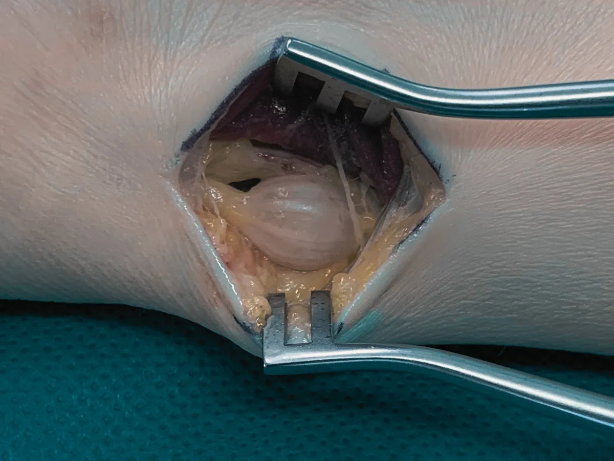
The Gold Standard for Schwannomas: Microsurgical Enucleation
Over time, the growth of a nerve tumor often leads to local swelling, pronounced tenderness to pressure, and radiating neuropathic pain, which is why surgical removal is usually the treatment of choice.
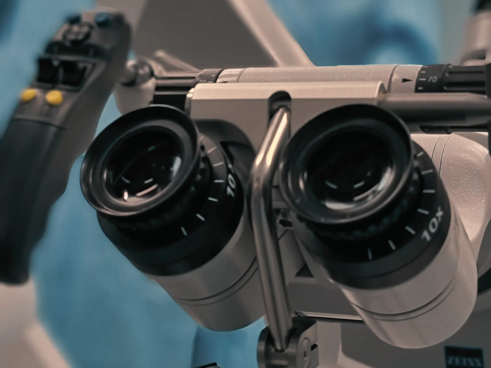
The absolute gold standard in the treatment of benign schwannomas is so-called microsurgical enucleation. Using specialized surgical microscopes, Dr. Megerle delicately opens the outer sheath of the nerve to carefully shell out the tumor from the inside.
To ensure the highest level of safety, we routinely use intraoperative neurophysiology (nerve stimulation). This allows us to electrically test individual nerve fibers during the ongoing surgery. This ensures that healthy, functional nerve tissue is accurately identified and preserved while the tumor is safely removed.
Frequently Asked Questions
What is a schwannoma (neurinoma)?
A schwannoma is a benign tumor that arises from Schwann cells, which form the protective sheath around peripheral nerves. They are the most common type of benign peripheral nerve tumor and typically present as well-defined, encapsulated growths.
How are peripheral nerve tumors diagnosed?
If a symptomatic peripheral nerve tumor is suspected, a high-resolution ultrasound or magnetic resonance imaging (MRI) is performed. These advanced imaging techniques help precisely locate the tumor and display its exact relationship to the functioning nerve fibers.
Will I suffer a loss of nerve function after the tumor is removed?
The primary goal of surgical treatment for a benign nerve tumor is complete removal while maximizing the preservation of motor and sensory functions. By using high-magnification surgical microscopes and continuous monitoring with intraoperative nerve stimulation, the tumor can be precisely separated from healthy, functioning nerve tissue, making permanent impairments rare.
Why are biopsies generally avoided for these tumors?
Closed biopsies (such as needle or core biopsies) are generally not recommended when a peripheral nerve tumor is suspected due to the significant risk of unintentional nerve damage. Instead, careful microsurgical exposure is the procedure of choice to maximally protect the nerve.